8SON | pdb_00008son

Crystal structure of macrophage migration inhibitory factor in complex with N-[3-(Trifluoromethyl)phenyl]-3-(2-chloroanilino)-2-cyano-3-thioxopropanamide


Experimental Data Snapshot

  • Method: X-RAY DIFFRACTION
  • Resolution: 1.63 Å
  • R-Value Free: 
    0.199 (Depositor), 0.210 (DCC) 
  • R-Value Work: 
    0.170 (Depositor), 0.180 (DCC) 
  • R-Value Observed: 
    0.171 (Depositor) 

Starting Model: experimental
View more details

wwPDB Validation   3D Report Full Report


Ligand Structure Quality Assessment 


This is version 1.1 of the entry. See complete history


Literature

Inhibition of MIF with an Allosteric Inhibitor Triggers Cell Cycle Arrest in Acute Myeloid Leukemia.

Pantouris, G.Khurana, L.Tilstam, P.Benner, A.Cho, T.Y.Lelaidier, M.Perree, M.Rosenbaum, Z.Leng, L.Foss, F.Bhandari, V.Verma, A.Bucala, R.Lolis, E.J.

(2025) ACS Omega 10: 17441-17452

  • DOI: https://doi.org/10.1021/acsomega.4c10969
  • Primary Citation of Related Structures:  
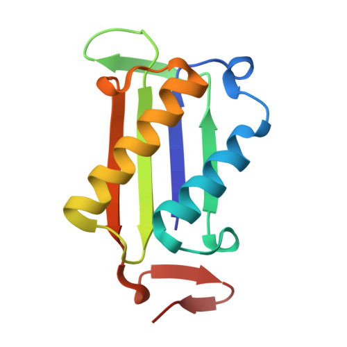
    8SON

  • PubMed Abstract: 

    Macrophage migration inhibitory factor (MIF) is a key modulator of innate and adaptive immunity that has been extensively reported to promote tumor cell survival, proliferation, and metastasis. A recent study focusing on the microenvironment of acute myeloid leukemia (AML) showed that pharmacological inhibition of MIF signaling, in vitro as well as in vivo , reduces AML cell survival. Such data highlights the crucial role of MIF in AML pathogenesis and support the efforts for developing selective MIF modulators. Here, we report the identification and crystallographic characterization of a MIF inhibitor (compound 1 ) with an allosteric binding motif. Single point screening of 1 against a panel of National Cancer Institute (NCI) 60 human tumor cell lines revealed a selective antitumor activity for the AML cell line HL-60. After confirming the protein's expression in multiple AML cell lines, we utilized 1 to extract mechanistic insights into MIF action. Our findings demonstrate that AML cells utilize an MIF-dependent proliferation mechanism, which upon inhibition triggers a G0/G1 cell cycle arrest of the malignant cells. Complementary analysis of the MIF receptors utilizing neutralizing antibodies and selective small molecule antagonists associates this effect with inhibition of CD74 activation. The collection of data presented herein highlights the important role of MIF in proliferation of AML cells and points to the need of developing small molecule anticancer therapeutics that target MIF signaling.


  • Organizational Affiliation

    Department of Chemistry, University of the Pacific, Stockton, California 95211, United States.


Macromolecules
Find similar proteins by:  (by identity cutoff)  |  3D Structure
Entity ID: 1
MoleculeChains Sequence LengthOrganismDetailsImage
Macrophage migration inhibitory factor
A, B, C
114Homo sapiensMutation(s): 0 
Gene Names: MIFGLIFMMIF
EC: 5.3.2.1 (PDB Primary Data), 5.3.3.12 (PDB Primary Data)
UniProt & NIH Common Fund Data Resources
Find proteins for P14174 (Homo sapiens)
Explore P14174 
Go to UniProtKB:  P14174
PHAROS:  P14174
GTEx:  ENSG00000240972 
Entity Groups  
Sequence Clusters30% Identity50% Identity70% Identity90% Identity95% Identity100% Identity
UniProt GroupP14174
Sequence Annotations
Expand
  • Reference Sequence
Small Molecules
Ligands 3 Unique
IDChains Name / Formula / InChI Key2D Diagram3D Interactions
30Y (Subject of Investigation/LOI)
Query on 30Y

Download Ideal Coordinates CCD File 
F [auth A],
I [auth B]
(2R)-3-[(2-chlorophenyl)amino]-2-cyano-3-thioxo-N-[3-(trifluoromethyl)phenyl]propanamide
C17 H11 Cl F3 N3 O S
JBTPUMQIJZGWRT-GFCCVEGCSA-N
SO4
Query on SO4

Download Ideal Coordinates CCD File 
E [auth A],
H [auth B],
K [auth C]
SULFATE ION
O4 S
QAOWNCQODCNURD-UHFFFAOYSA-L
GOL
Query on GOL

Download Ideal Coordinates CCD File 
D [auth A],
G [auth B],
J [auth C]
GLYCEROL
C3 H8 O3
PEDCQBHIVMGVHV-UHFFFAOYSA-N
Binding Affinity Annotations 
IDSourceBinding Affinity
30Y BindingDB:  8SON Ki: 1840 (nM) from 1 assay(s)
IC50: 3.31e+4 (nM) from 1 assay(s)
Experimental Data & Validation

Experimental Data

  • Method: X-RAY DIFFRACTION
  • Resolution: 1.63 Å
  • R-Value Free:  0.199 (Depositor), 0.210 (DCC) 
  • R-Value Work:  0.170 (Depositor), 0.180 (DCC) 
  • R-Value Observed: 0.171 (Depositor) 
Space Group: P 21 21 21
Unit Cell:
Length ( Å )Angle ( ˚ )
a = 68.534α = 90
b = 68.827β = 90
c = 89.329γ = 90
Software Package:
Software NamePurpose
REFMACrefinement
HKL-2000data reduction
PHASERphasing
PDB_EXTRACTdata extraction
HKL-2000data scaling

Structure Validation

View Full Validation Report


Ligand Structure Quality Assessment 


Entry History & Funding Information

Deposition Data


Funding OrganizationLocationGrant Number
Not funded--

Revision History  (Full details and data files)

  • Version 1.0: 2024-05-22
    Type: Initial release
  • Version 1.1: 2025-06-04
    Changes: Database references, Structure summary