8HKS | pdb_00008hks

Mutated human ADP-ribosyltransferase 2 (PARP2) catalytic domain bound to Pamiparib(BGB-290)


Experimental Data Snapshot

  • Method: X-RAY DIFFRACTION
  • Resolution: 2.80 Å
  • R-Value Free: 
    0.238 (Depositor), 0.240 (DCC) 
  • R-Value Work: 
    0.181 (Depositor), 0.180 (DCC) 
  • R-Value Observed: 
    0.184 (Depositor) 

Starting Model: in silico
View more details

wwPDB Validation   3D Report Full Report


Ligand Structure Quality Assessment 


This is version 1.1 of the entry. See complete history


Literature

Engaging an engineered PARP-2 catalytic domain mutant to solve the complex structures harboring approved drugs for structure analyses.

Wang, X.Zhou, J.Xu, B.

(2025) Bioorg Chem 160: 108471-108471

  • DOI: https://doi.org/10.1016/j.bioorg.2025.108471
  • Primary Citation of Related Structures:  
    8HKN, 8HKO, 8HKS, 8HLJ, 8HLQ, 8HLR

  • PubMed Abstract: 

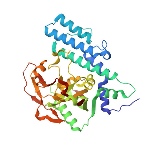
    The PARP-1/2 inhibitors have been approved for the treatment of cancers by modulating the enzymatic activity and/or the trapping ability for damaged DNA of PARP-1 and/or PARP-2, and the selective PARP-1 inhibitors are now attracting considerable attention with an aim to search for drug candidates with an improved safety. Exploring the structural basis of the selectivity and trapping capability of known PARP-1/2 inhibitors would be beneficial for the discovery of the improved inhibitors. Herein, a mutated PARP-2 catalytic domain, designated as catPARP-2SE, was engineered. It could be expressed in an elevated level and had capability to crystalize at 25 °C, which greatly facilitated obtaining PARP-2 crystals. Consequently, the complex structures of Fluzoparib, Pamiparib, Rucaparib, and Niraparib within PARP-2 were achieved. Taking advantage of these complexed structures, the detailed and quantitative analyses of protein-ligand and intra-protein interactions (αB-αF, αJ-αB, αJ-αF, ASL-αD and ASL-αF interfaces) were conducted with quantum chemistry methods (GFN2-xTB and IGMH). It suggested that the residues adjacent to Asp766 in the HD and ASL domains and the αJ-αF and ASL-αD interfaces were closely related to the selectivity and trapping mechanism. These results would provide some insights for the design and development of novel PARP-1/2 inhibitors with improved pharmacodynamic properties.


  • Organizational Affiliation

    Beijing Key Laboratory of Active Substances Discovery and Druggability Evaluation, Institute of Materia Medica, Chinese Academy of Medical Sciences and Peking Union Medical College, Beijing 100050, China.


Macromolecules
Find similar proteins by:  (by identity cutoff)  |  3D Structure
Entity ID: 1
MoleculeChains Sequence LengthOrganismDetailsImage
Poly [ADP-ribose] polymerase 2
A, B, C, D
353Homo sapiensMutation(s): 4 
Gene Names: PARP2ADPRT2ADPRTL2
EC: 2.4.2.30 (PDB Primary Data), 2.4.2 (PDB Primary Data)
UniProt & NIH Common Fund Data Resources
Find proteins for Q9UGN5 (Homo sapiens)
Explore Q9UGN5 
Go to UniProtKB:  Q9UGN5
PHAROS:  Q9UGN5
GTEx:  ENSG00000129484 
Entity Groups  
Sequence Clusters30% Identity50% Identity70% Identity90% Identity95% Identity100% Identity
UniProt GroupQ9UGN5
Sequence Annotations
Expand
  • Reference Sequence
Small Molecules
Ligands 2 Unique
IDChains Name / Formula / InChI Key2D Diagram3D Interactions
DS9 (Subject of Investigation/LOI)
Query on DS9

Download Ideal Coordinates CCD File 
G [auth A],
M [auth B],
P [auth C],
T [auth D]
(2R)-14-fluoro-2-methyl-6,9,10,19-tetrazapentacyclo[14.2.1.02,6.08,18.012,17]nonadeca-1(18),8,12(17),13,15-pentaen-11-one
C16 H15 F N4 O
DENYZIUJOTUUNY-MRXNPFEDSA-N
GOL
Query on GOL

Download Ideal Coordinates CCD File 
E [auth A]
F [auth A]
H [auth B]
I [auth B]
J [auth B]
E [auth A],
F [auth A],
H [auth B],
I [auth B],
J [auth B],
K [auth B],
L [auth B],
N [auth C],
O [auth C],
Q [auth D],
R [auth D],
S [auth D]
GLYCEROL
C3 H8 O3
PEDCQBHIVMGVHV-UHFFFAOYSA-N
Binding Affinity Annotations 
IDSourceBinding Affinity
DS9 BindingDB:  8HKS IC50: min: 0.11, max: 500 (nM) from 5 assay(s)
Experimental Data & Validation

Experimental Data

  • Method: X-RAY DIFFRACTION
  • Resolution: 2.80 Å
  • R-Value Free:  0.238 (Depositor), 0.240 (DCC) 
  • R-Value Work:  0.181 (Depositor), 0.180 (DCC) 
  • R-Value Observed: 0.184 (Depositor) 
Space Group: P 1 21 1
Unit Cell:
Length ( Å )Angle ( ˚ )
a = 81.006α = 90
b = 86.333β = 112.44
c = 103.619γ = 90
Software Package:
Software NamePurpose
autoPROCdata processing
MOLREPphasing
Aimlessdata scaling
PHENIXrefinement

Structure Validation

View Full Validation Report


Ligand Structure Quality Assessment 


Entry History & Funding Information

Deposition Data


Funding OrganizationLocationGrant Number
National Natural Science Foundation of China (NSFC)China82003657
National Natural Science Foundation of China (NSFC)China81673300
National Natural Science Foundation of China (NSFC)China82173693

Revision History  (Full details and data files)

  • Version 1.0: 2024-05-01
    Type: Initial release
  • Version 1.1: 2025-05-14
    Changes: Database references, Structure summary